Patient education
Forefoot Conditions
Neuroma
What is a Neuroma?
A neuroma is a painful condition caused by irritation and thickening of a nerve in the foot. It most commonly affects one of the small nerves that runs between the bones of the forefoot, usually between the third and fourth toes, but it can occur between other toes as well.
- Tight or narrow footwear
- High-heeled shoes that increase pressure on the forefoot
- Foot structure (such as flat feet or high arches)
- Abnormal foot mechanics during walking
- Repetitive impact activities (running, jumping, sports)
- Previous injury or trauma to the foot
What does a neuroma feel like?
Symptoms may include:
- Burning or sharp pain in the ball of the foot
- Tingling, numbness, or “electric shock” sensations in the toes
- Feeling like you are standing on a pebble or fold in your sock
- Pain that worsens with walking or tight shoes and improves when shoes are
removed
Symptoms often develop gradually and may worsen over time if untreated.
Simple summary
A neuroma is a thickened and irritated nerve in the forefoot that develops from
ongoing pressure or friction. It can cause pain, burning, or numbness between the
toes, especially when walking or wearing tight shoes. It is a common and treatable condition.
Bunion (Hallux Valgus)
What is a bunion?
A bunion (medical term: hallux valgus) is a structural deformity of the big toe joint
where the big toe gradually shifts toward the second toe and the bone at the base of the big toe becomes prominent on the inside of the foot.
This change in alignment affects the bones, ligaments, and muscles of the forefoot.
Over time, the joint may become inflamed, stiff, and painful.
A bunion is not just a bony lump — it is a progressive change in the position of the
joint and surrounding soft tissues.
What causes a bunion?
Bunions develop due to a combination of factors, including:
- Inherited foot shape and biomechanics
- Flat feet or unstable joints
- Abnormal walking patterns
- Tight, narrow, or poorly fitting footwear
- High-heeled shoes that increase pressure on the big toe joint
- Certain forms of arthritis
- Previous injury or trauma to the big toe joint
What are the symptoms?
- A visible bump on the inside of the big toe joint
- Pain or aching in the big toe joint
- Redness, swelling, or inflammation
- Stiffness or reduced movement of the big toe
- Difficulty fitting into shoes comfortably
- Callus or skin irritation from shoe rubbing
- Pain that worsens with walking or prolonged standing
Symptoms can range from mild to severe and may worsen over time.
How is a bunion diagnosed?
A bunion is diagnosed through:
- A clinical examination of your foot and walking pattern
- Assessment of joint movement and alignment
- Review of your symptoms and footwear
- X-rays, if needed, to measure bone alignment and assess joint health
Imaging helps determine the severity of the bunion and guide treatment options.
Non-surgical treatment options
Many bunions can be managed without surgery. Treatment may include:
- Footwear modification (wide toe-box shoes, low heels)
- Padding or protective dressings to reduce pressure
- Orthotics or insoles to improve foot mechanics
- Activity modification
- Anti-inflammatory strategies (if appropriate)
- Exercises to maintain joint mobility and strength
These treatments aim to reduce pain and slow progression but do not reverse the deformity.
Surgical treatment options
Surgery may be considered when:
- Pain persists despite appropriate non-surgical treatment
- Daily activities or footwear are significantly limited
- The deformity continues to worsen
Surgery aims to realign the bones, restore joint function, and relieve pain. There are many surgical techniques, and the most suitable procedure depends on: - Severity of the bunion
- Joint condition
- Foot structure
- Activity level
- Overall health
Your surgical podiatrist will discuss the most appropriate option for you.
Recovery after bunion surgery
Recovery varies depending on the procedure performed. General timelines may
include:
- Weeks 1-2: Wound care and reduced activity
- Weeks 3-4: Gradual return to walking and footwear
- Weeks 4-6: Return to normal activities
- Several months: Swelling continues to improve
Can bunions be prevented?
While bunions cannot always be prevented, you can reduce symptoms and slow progression by:
- Persistent pain in the big toe joint
- Difficulty walking or wearing shoes
- Increasing deformity
- Redness, swelling, or skin breakdown
- Numbness or tingling
Early management can help prevent worsening symptoms.
Summary
A bunion is a misalignment of the big toe joint where the toe drifts toward the second toe and a bony prominence forms on the inside of the foot. It may cause pain, swelling, and difficulty with footwear. Bunions are common and can often be
managed with conservative care, but surgery may be considered when symptoms
significantly affect daily life.
Digital Deformities (Hammer Toe, Mallet Toe & Claw Toes)
What are digital deformities?
Digital deformities are conditions where one or more toes bend or curl into an abnormal position due to an imbalance in the muscles, tendons, and joints of the
foot. Over time, these deformities can become stiff and fixed, making them painful
and difficult to manage with footwear.
The most common types of digital deformities are:
- Hammer toe
- Mallet toe
- Claw toes
Although they look similar, they affect different joints in the toe and have different
causes and treatments.
Understanding toe joints (simple anatomy)
Each toe has three small joints
- Metatarsophalangeal joint (MTPJ): where the toe meets the foot
- Proximal interphalangeal joint (PIPJ): the middle toe joint
- Distal interphalangeal joint (DIPJ): the end toe joint
Digital deformities occur when these joints bend abnormally due to muscle and tendon imbalance.
Types of digital deformities Hammer Toe
Hammer toe occurs when the middle joint of the toe (PIP joint) bends downward,
causing the toe to resemble a hammer shape.
Features:
- Bend at the middle of the toe
- Tip of the toe may point downward
- Often affects the second toe
- Can cause corns or calluses on the top of the toe
- May be flexible at first and become rigid over time
Mallet Toe
Mallet toe occurs when only the end joint of the toe (DIP joint) bends downward.
Features:
- Bend at the middle of the toe
- Tip of the toe may point downward
- Often affects the second toe
- Can cause corns or calluses on the top of the toe
- May be flexible at first and become rigid over time
Hammer toe occurs when the middle joint of the toe (PIP joint) bends downward,
causing the toe to resemble a hammer shape.
Features:
- at the middle of the toe
- Tip of the toe may point downward
- Often affects the second toe
- Can cause corns or calluses on the top of the toe
- May be flexible at first and become rigid over time
Mallet Toe
Mallet toe occurs when only the end joint of the toe (DIP joint) bends downward.
Features:
- at the middle of the toe
- Tip of the toe may point downward
- Often affects the second toe
- Can cause corns or calluses on the top of the toe
- May be flexible at first and become rigid over time
Claw Toes
Claw toes occur when the toe bends upward at the base joint (MTP joint) and
downward at both the middle (PIP) and end (DIP) joints, creating a claw-like
appearance.
Features:
- Toes appear clawed or curled
- Often affects multiple toes
- Frequently associated with nerve or muscle conditions
- Can cause painful pressure areas on the toes and ball of the foot
- May become rigid and difficult to straighten
What causes digital deformities?
- Toes appear clawed or curled
- Often affects multiple toes
- Frequently associated with nerve or muscle conditions
- Can cause painful pressure areas on the toes and ball of the foot
- May become rigid and difficult to straighten
Digital deformities develop due to muscle imbalance and pressure over time.
Contributing factors include:
- Tight or narrow footwear
- High-heeled shoes
- Flat feet or high arches
- Bunions or other foot deformities
- Long second toe
- Nerve or muscle disorders (more common with claw toes)
- Diabetes or neurological conditions
- Previous injury
- Age-related changes in soft tissues
What symptoms can occur?
Symptoms may include:
- Bent or curled toes
- Pain in the toes or ball of the foot
- Corns and calluses on the top or tip of the toes
- Skin irritation from shoe rubbing
- Difficulty fitting into shoes
- Redness or swelling
- Reduced toe movement
Symptoms often worsen over time if untreated.
How are digital deformities diagnosed?
Symptoms may include:
- Bent or curled toes
- Pain in the toes or ball of the foot
- Corns and calluses on the top or tip of the toes
- Skin irritation from shoe rubbing
- Difficulty fitting into shoes
- Redness or swelling
- Reduced toe movement
- Clinical examination of foot structure and toe position
- Assessment of flexibility (whether the toe can be straightened)
- Review of symptoms and footwear
- X-rays if needed to assess bone alignment and joint changes
Non-surgical treatment options
Early and flexible deformities may be managed with:
- Footwear changes (wide toe box, soft uppers)
- Padding or toe sleeves
- Orthotics or insoles to improve foot mechanics
- Toe spacers or splints
- Activity modification
- Corn and callus care
- Exercises and stretching
These treatments help relieve pressure and pain but do not permanently correct
fixed deformities.
Surgical treatment options
Surgery may be considered if:
- Pain persists despite conservative treatment
- The deformity becomes rigid
- There are recurrent corns, ulcers, or skin breakdown
- Footwear is very difficult to tolerate
Surgery aims to straighten the toe, rebalance tendons, and relieve pressure. The procedure chosen depends on:
- Type of deformity
- Flexibility of the toe
- Number of toes involved
- Overall foot structure
- General health
Recovery depends on the procedure but may include:
- First 1–2 weeks: swelling and wound care
- Weeks 3–6: walking in a surgical shoe or boot
- 6–8 weeks: gradual return to normal footwear
- Several months: swelling continues to improve
Your surgeon will guide you on weight-bearing and activity levels.
When should I seek professional advice?
You should seek assessment if you have:
- Painful or worsening toe deformities
- Recurrent corns or calluses
- Difficulty walking or wearing shoes
- Skin breakdown or ulcers
- Numbness or tingling
- Diabetes or circulation problems with toe deformities
Summary
Digital deformities occur when the toes bend into abnormal positions due to muscle and tendon imbalance.
- Hammer toe bends at the middle joint
- Mallet toe bends at the end joint
- Claw toes bend at multiple joints
They can cause pain, corns, and difficulty with footwear. Many cases can be
managed with non-surgical care, but surgery may be considered if symptoms
significantly affect daily life.
Warts
What Are Warts?
Warts are small, rough growths on the skin caused by the human papillomavirus
(HPV). They can appear anywhere on the body but are most common on the hands, feet, and fingers. Warts are generally harmless, but they can be unsightly,
uncomfortable, or sometimes painful, especially on weight-bearing areas like the soles of the feet.
Treatment Options
There are several effective treatments available for warts, depending on their size, location, and severity:
1. Topical Treatments – These include creams, gels, or solutions that contain
active ingredients such as salicylic acid. They work by softening the wart over
time so it can gradually be removed. This method may take several weeks of
consistent use.
2. Cryotherapy (Freezing) – In this treatment, the wart is frozen using liquid
nitrogen, causing it to blister and eventually fall off. Multiple sessions may be
needed for stubborn warts.
3. Curettage (Scraping/Excision) – Curettage is a quick, in-clinic procedure
performed under local anesthesia. The wart is carefully scraped off using a
small surgical instrument called a curette, often after softening the area with a
chemical or freezing treatment.
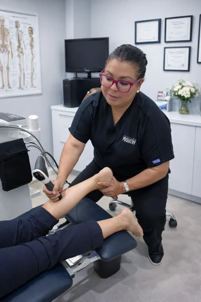
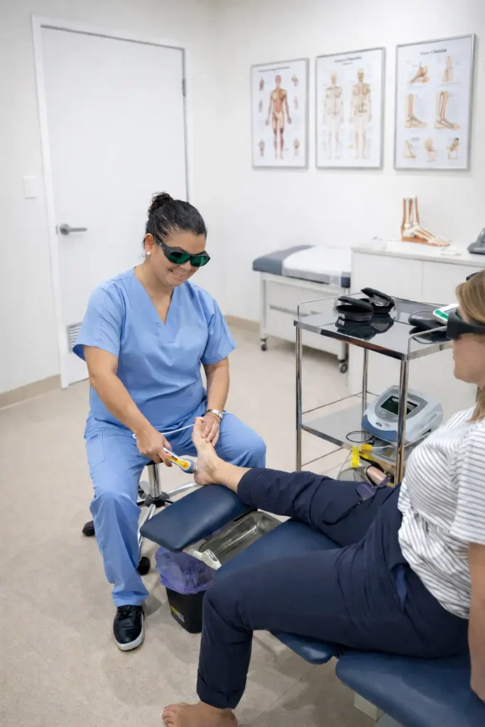
- How it works: The surgical podiatrist carefully removes the wart tissue layer, allowing for thorough clearance. This is undertaken generally under local anaesthesia.
- What to expect: Following administration of local anaesthetic, the procedure is typically well tolerated with little to no discomfort during treatment. As the wart is removed, a small superficial wound remains, which may be prone to minor bleeding. A firm, supportive dressing is applied to provide compression and minimise this. The area may be tender for a few days, with healing generally occurring over 1–2 weeks. The patient will be provided with clear aftercare instructions to support healing, reduce the risk of infection, and promote optimal tissue recovery with minimal scarring.
- Advantages: Curettage is a quick and effective treatment option, particularly for warts that have not responded to conservative treatment options.
Preventing Warts
- Avoid direct contact with warts on other people or on yourself
- Keep your skin clean and dry
- Avoid walking barefoot in public areas like pools or communal showers
- Do not pick or scratch warts, as this can spread the virus
Toe Fractures
A fractured toe is a common injury that occurs when one of the small bones in the toe is broken. The most frequent causes include dropping a heavy object on the foot, stubbing the toe, or twisting the foot.
Symptoms
Symptoms of a fractured toe may include:
- Pain, swelling, and tenderness at the site of injury
- Bruising or discoloration
- Difficulty walking or putting weight on the foot
- Deformity or crooked appearance in some cases
If you suspect a fractured toe, it’s important to see your healthcare provider for an evaluation. Diagnosis usually involves:
- Clinical examination, assessing pain, swelling, and deformity
- Imaging, such as X-rays, to confirm the break and assess alignment
Treatment Options
Treatment depends on the severity and type of fracture:
- Rest and Immobilisation
- The affected toe may be taped to an adjacent toe (buddy taping) or placed in a splint or cast to limit movement and promote healing.
- Protective footwear may also be recommended.
2. Ice and Elevation
- Applying ice and keeping the foot elevated helps reduce swelling and pain.
3. Pain Management
- Over-the-counter medications such as paracetamol or ibuprofen can help manage discomfort.
4. Surgery
- Severe fractures, displaced bones, open fractures, or fractures involving the joint may require surgical realignment and fixation using pins, screws, or small plates.
5. Follow-Up Care
- Some fractures may require physical therapy to restore range of motion, strength, and function to the affected foot.
Recovery
- Healing generally takes 4–8 weeks for uncomplicated fractures
- Weight-bearing may be restricted initially, with gradual return to normal activity
- Prompt evaluation and proper care are essential to prevent complications and ensure proper healing
Soft tissue & structural lumps
Ganglion (ganglion cyst)
- On the top of the foot
- Around the ankle
- Near toe joints
- Along tendon sheaths
- Repetitive stress or overuse of a joint or tendon
- Joint or tendon irritation
- Previous injury or trauma
- Weakness in the joint capsule or tendon sheath
- Arthritis or joint degeneration
- Adults and adolescents
- Active individuals and athletes
- People who spend long periods standing or walking
- Those with previous foot or ankle injuries
- A visible or palpable lump under the skin
- Localised swelling
- Aching or discomfort
- Pain with footwear pressure
- Tingling or numbness if the cyst presses on a nerve
- Changes in size (may get larger or smaller over time)
Some ganglions cause no pain and are noticed only as a lump.
- Clinical examination of the lump
- Review of symptoms and footwear irritation
- Assessment of joint and tendon movement
- Ultrasound to confirm it is fluid-filled
- MRI if the diagnosis is uncertain or to assess deeper structures
- X-rays to rule out bone-related problems
- Observation and monitoring
- Footwear modification to reduce pressure
- Padding or protective dressings
- Activity modification
- Orthotics if foot mechanics contribute to irritation
- Aspiration (draining the fluid with a needle) in selected cases
- The ganglion is painful or growing
- It interferes with walking or footwear
- It compresses a nerve
- Conservative treatment has failed
- The diagnosis is uncertain
- Weeks 1-2: Wound care and reduced activity
- Weeks 3-4: Gradual return to walking and footwear
- Weeks 4-6: Return to normal activities
- Several months: Swelling continues to improve
Can ganglions be prevented?
- Wearing well-fitting shoes
- Avoiding excessive pressure over joints
- Managing foot biomechanics with orthotics if recommended
- Treating injuries promptly
- Avoiding repeated trauma to the same area
- A growing or painful lump
- Difficulty wearing shoes
- Numbness or tingling
- Skin changes over the lump
- A lump that does not improve
- Any uncertainty about the diagnosis
A ganglion is a benign, fluid-filled lump that develops near a joint or tendon in the foot or ankle. It may cause pain or discomfort depending on its size and location. Many ganglions can be managed without surgery, but surgery may be recommended if symptoms persist or interfere with daily activities.
Plantar fibroma (plantar fibromatosis)
- Repetitive strain or micro-trauma to the plantar fascia
- Genetic or family tendency
- Tight calf muscles or plantar fascia
- Flat feet or high arches
- Previous foot injury
- Certain medical conditions (such as diabetes or epilepsy)
- Some medications
Who gets plantar fibromas?
- Adults more commonly than children
- Men more often than women
- People who spend long periods standing or walking
- Athletes and active individuals
- People with a family history of fibromas
- A firm lump or nodule in the arch of the foot
- Pain or discomfort when walking or standing
- Tenderness when pressure is applied
- A feeling of “walking on a pebble”
- Pain that worsens with tight shoes or barefoot walking
- Sometimes no pain at all (the lump is noticed before symptoms)
How is a plantar fibroma diagnosed?
Diagnosis is based on:
- Clinical examination of the foot
- Assessment of the size, location, and firmness of the lump
- Review of symptoms and walking pattern
- Ultrasound to confirm a solid fibrous mass
- MRI for larger or unclear lumps
- X-rays to rule out bone-related problems
Most plantar fibromas are managed without surgery. Treatment may include:
- Padding or cushioning to reduce pressure
- Footwear modification (soft soles, wider shoes)
- Orthotics or insoles to offload the area
- Activity modification
- Stretching exercises for the plantar fascia and calf muscles
- Anti-inflammatory strategies if appropriate
- Corticosteroid injections in selected cases to reduce pain and size
Surgery may be considered when:
- Pain persists despite conservative treatment
- The fibroma interferes with walking or daily activities
- The lump continues to grow
- Footwear becomes difficult to tolerate
- Weeks 1-2: Wound care and limited weight-bearing
- Weeks 3-6: Gradual return to walking in supportive footwear
- Weeks 6-8: Increasing activities
- Several months: Full healing and scar maturation
- Wearing supportive, well-fitting footwear
- Using orthotics if recommended
- Stretching the calves and plantar fascia
- Avoiding repeated trauma to the arch of the foot
- Treating foot pain early
- A new lump in the arch of your foot
- Increasing pain or tenderness
- Difficulty walking or standing
- Rapid growth of the lump
- Skin changes over the area
- Any uncertainty about what the lump is
Arthritis & joint degeneration
Hallux Rigidus / Hallux Limitus
Hallux rigidus is the second most common disabling deformity of the first metatarsophalangeal (MTP) joint, second only to hallux valgus. It is characterised by pain and limited motion at the big toe joint, often affecting daily activities such as walking, running, or wearing shoes comfortably.
Terminology
Hallux limitus is distinct from hallux rigidus. While hallux rigidus refers to structural osteoarthritis of the first MTP joint, hallux limitus describes functional pain secondary to limited flexibility of surrounding soft tissues, such as gastrocnemius spasm. Despite this difference, hallux limitus is considered a risk factor for the development of hallux rigidus, and features of both conditions can co-occur.
Epidemiology Hallux rigidus is most observed in middle-aged adults, though it can occasionally develop during adolescence. Unlike hallux valgus, it appears to slightly affect males more than females.
Diagnosis
Diagnosis is based on:
- Pain localised to the first MTP joint
- Clinical findings of decreased dorsiflexion (<30°)
- Palpable osteophytes at the joint margins
Patients typically present with pain at the big toe joint, often worsened by walking or wearing shoes. The functional disability from hallux rigidus is generally greater than that seen in hallux valgus, as dorsiflexion of the joint is severely restricted and painful. Removing shoes does not relieve the discomfort.
Radiographic Features
The hallmark of hallux rigidus is osteoarthritis of the first MTP joint, which can be seen across various imaging modalities. Additional findings may include:
- Widening of the first metatarsal head
- Associated hallux valgus, although its significance is debated
- Metatarsus primus elevatus, or relative dorsal elevation of the first metatarsal, which may be causative or secondary
Classification
Hattrup and Johnson Radiographic Classification:
- Grade 1: Mild to moderate osteophyte formation with good joint space preservation
- Grade 2: Moderate osteophyte formation, joint space narrowing, and subchondral sclerosis
- Grade 3: Marked osteophyte formation, loss of visible joint space, ± subchondral cysts
Coughlin and Shurnas Functional & Radiographic Classification:
- Grade 0: Dorsiflexion 40–60°, normal radiograph, no pain
- Grade 1: Dorsiflexion 30–40°, dorsal osteophytes, minimal joint changes
- Grade 2: Dorsiflexion 10–30°, mild to moderate joint narrowing or sclerosis, presence of osteophytes
- Grade 3: Dorsiflexion <10°, severe radiographic changes, constant moderate to severe pain
- Grade 4: Stiff joint, severe degenerative changes with loose bodies and osteochondritis dissecans
Treatment and Prognosis
Treatment depends on severity, symptoms, and functional impact:
- Conservative management may include footwear modifications, orthotics, anti-inflammatory medications, and activity adjustment.
- Surgical options include:
-
- Osteophyte resection (cheilectomy) and capsular release – for early-stage disease with preserved joint space
- Arthrodesis (joint fusion) – often the preferred option for advanced arthritis, providing pain relief and stability
- Arthroplasty (joint replacement) – considered in select patients to maintain some range of motion
1st MTPJ arthrodesis
What is 1st MTPJ Arthrodesis?
1st MTPJ arthrodesis is a surgical procedure that fuses the joint at the base of the big toe. This joint is where the first metatarsal bone meets the proximal phalanx of the toe. Fusion is usually recommended for patients with severe arthritis, hallux rigidus (stiff big toe), deformity, or chronic pain that has not improved with conservative treatments.
The goal of 1st MTPJ arthrodesis is to relieve pain, improve function, and correct deformity in the big toe. It is typically considered when:
- Arthritis has caused severe joint degeneration
- The big toe has limited movement or causes pain while walking
- Previous treatments such as medications, orthotics, or injections have not been effective
What to Expect from the Procedure
During the surgery, the damaged cartilage is removed, and the bones are positioned to create a stable, pain-free joint. Small screws, plates, or pins are often used to hold the bones together while they fuse over several weeks.
Why is it done?
The goal of 1st MTPJ arthrodesis is to relieve pain, improve function, and correct deformity in the big toe. It is typically considered when:
- Arthritis has caused severe joint degeneration
- The big toe has limited movement or causes pain while walking
- Previous treatments such as medications, orthotics, or injections have not been effective
What to Expect from the Procedure
During the surgery, the damaged cartilage is removed, and the bones are positioned to create a stable, pain-free joint. Small screws, plates, or pins are often used to hold the bones together while they fuse over several weeks.
Recovery
- Recovery usually involves immobilisation in a special boot or cast for 6–8 weeks to allow the bones to heal
- Weight-bearing may be limited initially, and gradual physiotherapy can help restore mobility and strength
- Most patients experience significant pain relief and improved function, though the big toe will no longer bend at the fused joint
Benefits
- Long-term pain relief from arthritis
- Correction of deformity (e.g., bunions or misalignment)
- Improved stability and walking comfort
Considerations
- The loss of motion at the big toe is permanent, so patients need to understand the trade-off between pain relief and flexibility
- Some swelling, stiffness, or discomfort may persist for several months
As with any surgery, there are risks of infection, delayed healing, or hardware issues, which your surgeon will discuss.
Foot arthritis (general)
What is Foot Arthritis?
Foot arthritis occurs when the joints in the foot become inflamed and degenerate, leading to pain, stiffness, and reduced mobility. The condition can affect any joint in the foot, but most commonly impacts the big toe (first metatarsophalangeal joint), midfoot, and ankle joints. Arthritis may develop due to age-related wear and tear (osteoarthritis), previous injuries, or inflammatory conditions such as rheumatoid arthritis.
Symptoms
Common signs of foot arthritis include:
- Persistent joint pain or aching
- Swelling and tenderness around the joint
- Stiffness, particularly in the morning or after periods of rest
- Difficulty walking or wearing shoes comfortably
- Deformities such as bunions or joint misalignment in severe cases
Treatment Options
Treatment depends on the severity of arthritis and impact on daily life.
1. Conservative Management
- Rest and activity modification to reduce stress on affected joints
- Supportive footwear and orthotics to improve alignment and reduce pressure
- Anti-inflammatory medications for pain relief
2. Cortisone Injections
- Cortisone is a powerful anti-inflammatory medication that can be injected directly into the affected joint
- Injections help reduce pain and swelling, providing temporary relief and improving mobility
- The effects can last weeks to months, and injections may be repeated if necessary
3. Surgery
- When conservative measures and cortisone injections no longer provide sufficient relief, surgery may be recommended
- Surgical options vary depending on the joint affected and severity of arthritis and may include:
- Arthrodesis (joint fusion) – stabilizes the joint and relieves pain by fusing the bones
- Joint replacement – replaces the damaged joint with an artificial implant to restore function
- Osteotomy or realignment procedures – correct deformities caused by arthritis
- Surgery aims to reduce pain, restore function, and improve walking comfort, though recovery can take several weeks to months
- Surgical options vary depending on the joint affected and severity of arthritis and may include:
When to See a Surgical Podiatrist
If foot pain is persistent, worsening, or affecting daily activities, it’s important to seek professional assessment. Early, targeted intervention can help manage symptoms, maintain joint function, and preserve mobility. While some treatments may also slow disease progression—particularly in inflammatory forms of arthritis—this is less predictable in degenerative conditions such as osteoarthritis.
Ready to take the next step?
Whether you need a consultation or are ready for treatment, we’re here to help you get back on your feet.